Breaking the Code: How AI Cheating is Transforming College Campuses
Artificial intelligence (AI) has come a pretty long way from its earliest iterations. Large language models (LLMs) have "learned" a lot about what works and what doesn’t in academia. However, this has become a problem for many college lecturers and teaching assistants. A significant question comes to light in these considerations: Is using AI cheating?
Read MoreLatest News


Trending Stories


Self-introduction is an inevitable part of daily interactions within your social, academic, and professional circles. As we transition to new neighborhoods, colleges, and workplaces, we have to int...
Admission


StudyTech


The five-paragraph essay format is a simple structure that helps the writer organize their thoughts into five paragraphs. Because of its structure, it's also known as a hamburger essay, one three o...
Student Life


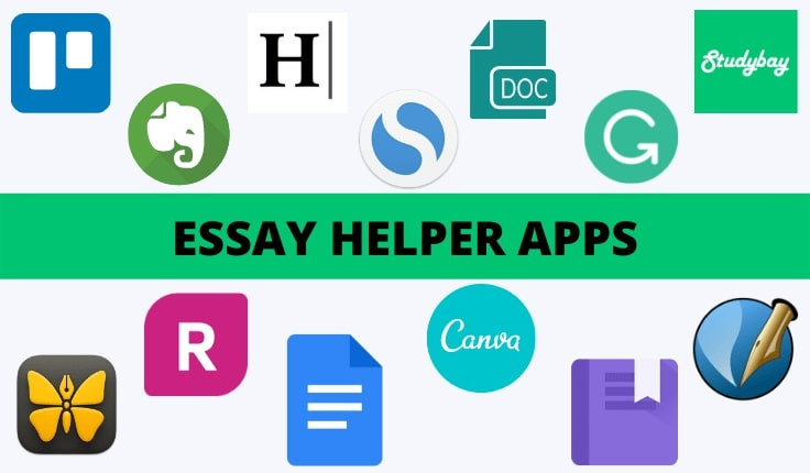
There are hundreds of essay apps available, with purposes ranging from note-taking and planning to checking grammar. To help you choose the best app for your essay, we’ve created a comprehensive li...